What happens as you’re reading this article? When you see the look on your mom’s face after you come home late at night? When you stare up at the sunset after a long day at work, and find yourself rather relaxed? You’re utilizing vision to the maximum potential!! And, I don’t think it’s a secret that our visual system is amongst the most unique and complex systems of our body. It utilizes potentially the smallest entity in the world, one measly (yet oddly speedy) particle of light, in order to craft a wondrous image of sheer beauty. While that mechanism in and of itself is absolutely fascinating, it masks an often underrated piece of vision…the way we tell things apart. Off the top of our heads, it seems simple, right? Different colors, different shapes, blah blah blah. But, in reality, it’s a process which is a lot more exciting…in the literal sense, than you may think
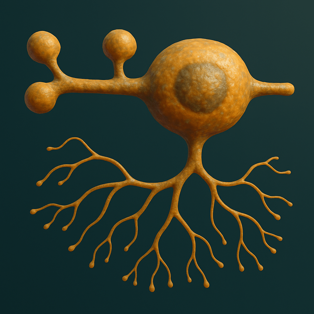
This tiny cell can do a lot, but that’s a pretty cliché way of magnifying the wondrous nature of biology. To be more specific, this tiny cell is quite literally responsible for us allowing us to distinguish between the objects surrounding us
Pieces Of The Puzzle
See The Light
As always, there’s no point in delving right into the intricate mechanisms of our systems if we do not have a functional understanding of the generalized physiology behind the entire process. Vision is complex, but we can break it down into elementary components, specifically its onset. As a photon strikes our eyes, it is absorbed by a specific type of cell, known as a photoreceptor (the nomenclature makes sense). If you’re somewhat versed in the behavior of the neural circuit, you’ll predict the course of action that is to follow. There is an ensuing action potential which follows within the photoreceptor, but it’s not like the standard action potential, where ionic channels will immediately open. The photoreceptor operates via a G-Protein Cascade, which sounds intimidating, but is simply scientific jargon which indicates that protein modifications down a sequence amplify an initial signal or onset (in this case, photon absorption). It is the conformational change that a class of molecules known as photopigments undergo which trigger sequence amplification and lead to the neuron being electrically stimulated down its chain, to then produce light. However, while you may be familiar with mechanisms of electrical stimulation for general neurons, we’ve got to redefine what reversing polarity actually means when it comes to photoreceptors

The photoreceptor is an extremely unique molecule. The functional protein behind its mechanism of action is known as a photopigment, which contains two components. ‘Retinal,’ which changes shape after absorbing a Photon, and ‘Opsin,’ whose response to the Retinal change determines which wavelengths that specific photoreceptor will respond to
Opposite Day
It’s a bit of a cliffhanger to suggest that photoreceptors literally ‘reverse polarity’ differently than all other neurons. And, it’s somewhat untrue. Reversing the polarity of an entity just indicates that the overall electrical charge present in an entity is now of an opposite sign (negative to positive, vice versa). However, the statement isn’t entirely false, in terms of the manner in which it happens. Neurons normally maintain a negative resting potential, and upon stimulation, have a positive electrical current flowing through them, leading to an entire elicited signal. Photoreceptors, however, are, at rest, relatively more positive than the average neuron. Note, this doesn’t mean that they are constantly stimulated; it just is a result of a constant cationic flow during the resting state. The G-Protein Cascade that eventually results in vision triggers a conformational change in cGMP, a protein that keeps these cationic channels open. Hence, the photoreceptor’s change to a more negative state, contrary to other neural mechanisms, produces a stimulus
The photoreceptor mechanism is, inherently, quite fascinating. Yes, it’s definitely interesting how they can operate via hyperpolarization, rather than depolarization…however, it’s not our job to determine whether it’s ‘cool,’ or not. It’s our job to determine the biological significance of such a feature
To The Side
Most of us in neuroscience are used to the traditional ‘down the line’ propagation. An action potential is fired in one neuron, leading to another neuron, and another, and another, until finally, a stimulus is produced. However, the visual system includes side components…in an absolutely literal sense. Before outlining the physiological reason for these ‘side components,’ we have to first understand the path that the visual current takes. From the photoreceptor, the current goes to Bipolar Cells, and then to Ganglion Cells, in a path to the Visual Cortex. However, there is a component I intentionally did not mention: Horizontal Cells. The reason I did not mention them as part of the general visual circuit is because technically, they are only modulators of the signal, and not cells that the signal directly propagates through. This group of cells are, however, an integral part of the visual system, precisely regulating the contents of what we see in a process deemed ‘Lateral Inhibition,’ a process which we will devote the rest of the article to discussing in depth
Side Characters
Moving Parts
While we tend to appreciate the circuit that leads to sight itself, the circuit would be completely haywire without a key visual feature known as contrast…a feature which quite literally allows us to separate light from dark. So, a key component of the visual system that must be understood is that Photoreceptors are arranged in a Center-Surround Model, and this denotation has everything to do with the placement of the Bipolars relative to the Photoreceptors. Those Photoreceptors which are connected to Bipolars are known as ‘Center,’ while the rest are surround. However, all photoreceptors in the field are connected to Horizontal Cells, which provide inhibitory feedback to Photoreceptors. However, as we know…inhibition supports photoreceptors. But, before I explain the actual mechanisms surrounding Lateral Inhibition, I have to detail a crucial nuance about Bipolars. Bipolars which are stimulated and produce a light based response are known as ‘ON’ Bipolars. Those which produce a response when exposed to decreased light are known as ‘OFF’ Bipolars. While this fact seems insignificant in the context of Horizontal Cells, it will allow us to gain an understanding of what sort of response a Horizontal Stimulus will lead to…and now, we can continue our discussion of the intricacies of lateral inhibition
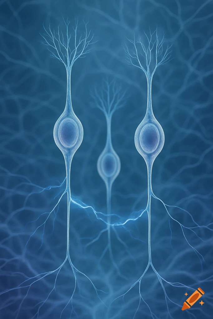
The image to the left is actually a rather accurate representation of a Bipolar Cell, named accordingly as there is only one branch extending from the Soma on either side: The Dendrite and The Axon. It is appropriately shaped, for it has no physiological need outside of propagating the visual signal to the Ganglion Cell
High Contrast Mode
The visual field operates entirely on this concept of Contrast. Without it, everything would likely appear distorted…no detection of edges, no differentiation between surfaces, blended colors, and such. Note, in all of the following scenarios, assume that the Photoreceptor connects to an ‘ON’ Bipolar. Consider a spot of light focused on the Center Photoreceptor, but dark on the Surround Cells. In this case, Glutamate is rich in these Surround Photoreceptors and the Horizontal Cells (who do follow the standard pattern of Glutamate Excitation) provide maximal inhibition to the Photoreceptors, as all besides a few are depolarized. Hence, the hyperpolarization of the Center Cone is further bolstered and light detection is quite enhanced, separating the light from the surrounding dark. However, what if light is encapsulating the whole receptive field? The entire field is hyperpolarized and Horizontals don’t provide much inhibitory feedback…there’s no contrast enhancement needed, for the whole field is lit up. Now, what if the Center Cone is not illuminated, despite Surround illumination. Horizontal inhibition isn’t prominent, as most of the field is hyperpolarized; there is no illumination enhancement on the Center, so the Dark Center is in thorough contrast to the Illuminated Surround. It is this simple yet illustrious visual mechanism that quite literally allows us to maximally separate the light…and the dark.
WRAPPING IT UP
Just when you thought it couldn’t get any more nuanced, neurobiology always has more surprises in store for us…some we likely haven’t even discovered yet!! But, such is where the beauty of science lies: The infinite scavenge for knowledge!


Leave a comment